Publikationen

Link zu aktuellen Büchern

Link zu Austrian Studies in Social Anthropology

Link zu den Vienna Working Papers in Ethnography

Link zu Journal of New Zealand and Pacific Studies

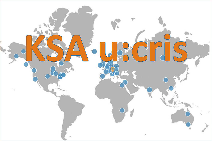
peer-reviewed Publikationen in u:cris

3D visualization of bioerosion in archaeological bone

Autor(en)
Kellie Sara Duffett Carlson, Kirsten Mandl, Ashley McCall, David Brönnimann, Maria Teschler-Nicola, Estella Weiss-Krejci, Brian Metscher
Abstrakt

Palaeoradiology is increasingly being used in archaeological and forensic sciences as a minimally invasive alternative to traditional histological methods for investigating bone microanatomy and its destruction by diagenetic processes. To better understand ancient mortuary practices, taphonomic studies using microCT scanning methods are gaining an ever more important role. Recently it was demonstrated that 2D virtual sections obtained by microCT scanning of intact samples are comparable to physical sections for the rating and diagnosis of bioerosion in archaeological bone. Importantly, volume image data obtained from tomographic methods also allow the rendering and analysis of 3D models. Building on these methods we provide (1) detailed descriptions of bioerosion in 3D volume renderings, virtual sections, and traditional micrographs, and (2) accessible techniques for the visualization of bioerosion in skeletal samples. The dataset is based on twenty-eight cortical bone samples, including twenty femora (of which five are cremated), two ribs, two parietals, one mandibular ramus, one humerus, and two faunal long bones from five archaeological sites in Lower Austria dating from the Early Neolithic to the Late Iron Age. Notably, we reduce the need for time-consuming image segmentation by sequentially applying two noise-reducing, edge-preserving filters, and using an image-display transfer function that visualizes bioerosion, as well as Haversian and Volkmann canal structure and density in 3D. In doing so we are also able to visualize in 3D the invasion of canals by microbiota, which has previously only been reported in 2D sections. Unlike conventional thin sections, the 3D volume images shown here are easy to create and interpret, even for archaeologists inexperienced in histology, and readily facilitate the illustration and communication of microtaphonomic effects.

Organisation(en)
Department für Evolutionäre Anthropologie, Institut für Urgeschichte und Historische Archäologie, Institut für Kultur- und Sozialanthropologie, Department für Evolutionsbiologie
Externe Organisation(en)
Österreichische Akademie der Wissenschaften (ÖAW), Universität Basel, Naturhistorisches Museum Wien (NHM), Independent researcher
Journal
Journal of Archaeological Science
Band
145
ISSN
0305-4403
DOI
https://doi.org/10.1016/j.jas.2022.105646
Publikationsdatum
09-2022
Peer-reviewed
Ja
ÖFOS 2012
106056 Biologische Anthropologie, 601025 Experimentelle Archäologie
Schlagwörter
ASJC Scopus Sachgebiete
Archaeology, Archaeology
Link zum Portal
https://ucrisportal.univie.ac.at/de/publications/3d-visualization-of-bioerosion-in-archaeological-bone(06202942-94fd-49d2-886d-74d6bb732091).html